Keyword [ChestX-ray14]
Wang X, Peng Y, Lu L, et al. Chestx-ray8: Hospital-scale chest x-ray database and benchmarks on weakly-supervised classification and localization of common thorax diseases[C]//Proceedings of the IEEE conference on computer vision and pattern recognition. 2017: 2097-2106.
1. Overview
1.1. Motivation
- there is still significant room for performance improvement when underlying challenges become greater (0.413 on COCO vs 0.884 on VOC)
In this paper, it present a new chest X-ray database (ChestX-ray8)
- comprise 108,948 frontal-view X-ray images of 32,717 unique patients
- with the text-mined 8 disease image labels
- 24,636 images contain one or more pathologies
- 84,312 images are normal cases, label [0, 0, 0, 0, 0, 0, 0, 0]
- resize image dimension from 3000x2000 (typical for X-ray image) to 1024x1024 bitmap

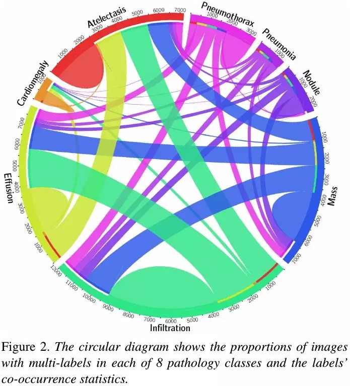
- some connection between different pathologies, which agree with radiologists’s domain knowledge, such as Infiltration is often associated with Atelectasis and Effusion
1.2. DCNN Framework

- exploit Global Average Pooling to visualize the region
1.2.1. Log-Sum-Exp (LSE) Pooling

- serve as an adjustable option between max pooling (r→ ∞) and average pooling (r→ 0)
- LSE suffers from overflow/underflow problems, so

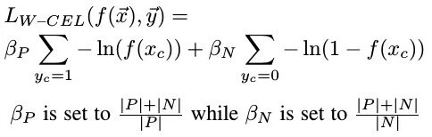
1.3. Loss Function
one of 3 standard loss function
- Hinge Loss (HL)
- Euclidean Loss (EL)
- Cross Entropy Loss (CEL)
1.3.1. Sample-Balance
- W-CLE
2. Experiments
2.1. Different Pooling Strategy

- r=10 is best
2.2. Comparison

- W-CEL is better than CEL